low flow low gradient aortic stenosis ase
13 3A The current diastolic profile see Figs. Jets low in momentum homogeneous in color aliasing mostly confined to the base of the jet.

Low Flow Low Gradient Severe Aortic Stenosis Despite Normal Ejection Fraction Is Associated With Severe Left Ventricular Dysfunction As Assessed By Speckle Tracking Echocardiography Circulation Cardiovascular Imaging
High-pressure gradient and high-velocity flow eg VSD Low pitch.

. Aortic stenosis is tightening of the aortic valve and mild aortic stenosis is a mild tightening. Mild Aortic Stenosis. Aortic stenosis with severe left ventricular dysfunction and low transvalvular pressure gradients.
American Society of Echocardiography 2100 Gateway Centre Boulevard Suite 310 Morrisville NC. Stress echocardiography with the addition of coronary flow velocity reserve measurement may be reasonable to improve diagnosis of coronary myocardial. Severe symptomatic aortic stenosis.
Chest pain of pericarditis increases in the supine position and may be associated with a friction rub. Valvular stenosis as the predominant lesion before the operation. Low-Risk Patients With Acute Chest Pain e392 4111.
Since the Intersocietal Accreditation Commission mandates both the assessment and reporting of diastolic function for echo accreditation we thought it would be a good idea to review these measurements again. Aortic stenosis is most commonly caused by age-related progressive calcification 50 of cases with a mean age of 65 to 70 years. New classification of AS by gradient flow and ejection fraction.
This pressure gradient develops immediately after the aortic valve closes which marks the start of diastole and the left ventricle starts to relax. Jander N Minners J Holme I et al. Low-flow states diagnosis management and a review of the current literature.
A comprehensive literature search was conducted from November 11 2017 to May 1 2020 encompassing studies reviews and other evidence conducted on human subjects that were published in English from PubMed EMBASE the Cochrane Collaboration Agency for Healthcare Research and Quality reports and other relevant databases. Challenges in aortic valve stenosis. Sherwood MW Kiefer TL.
In patients with low systolic function this can be further corroborated with an abnormal flow propagation Vp on color M-mode and a EVp greater than 2. Zoghbi MD FASE MACC Professor and Chairman Department of Cardiology Elkins Family Distinguished Chair in Cardiac Health Houston Methodist Hospital Flow dependence of. 164 x 085 1394.
Role of 2D3D and CMR Dobutamine Stress testing In Low Flow Low EF Low Gradient Aortic Stenosis Case Studies William A. Etiologies include congenital bicuspidunicuspid calcific and rheumatic disease. Generally in mild aortic stenosis the tight valve remains greater than 15-2 cm 2.
Another major cause of aortic stenosis is the calcification of a congenital bicuspid aortic valve or more rarely a congenital unicuspid aortic valve. Concentric LVH is the result of the heart adapting to high systemic pressure overload caused by hypertension or other diseases such as aortic stenosis. As we said above the usual aortic valve area is like a medium sized wristwatch around 3-4 cm 2 in area.
48 Likes 2 Comments - College of Medicine Science mayocliniccollege on Instagram. Stress echocardiography used as a screening test for ischemic heart disease in a patient without signs or symptoms is not covered. PE may result in tachycardia dyspnea and accentuated P2.
ME imaging of the AV and root. The New ASE Guidelines. This flow is propelled by the pressure gradient between the left atrium and the left ventricle.
Low Flow Low Gradient AS with Reduced Ejection Frac-tion 384 Low Flow Low Gradient AS. 狭窄Low flow low gradient AS with preserved ejection fraction. J Am Coll Cardiol.
Low-dose dobutamine may be useful for assessing for low-gradient AS. Noncoronary causes of chest pain include aortic stenosis aortic regurgitation and hypertrophic cardiomyopathy which produces characteristic murmurs and pulse alterations. Concentric LVH affects both men and women regardless of age.
If your image is chosen you will receive an American Society of Echocardiography guideline poster of your choice and be recognized in a subsequent CASE issue. Dobutamine stress echo may be indicated to detect low gradient low output aortic stenosis or clinically silent transplant coronary disease. Severe systemic arterial hypertension eg 200110 mm Hg.
Interestingly this means the valve. The E-wave represents passive blood flow from the left atrium to the left ventricle. Choosing the Right Pathway With Patient-Centric Algorithms for Acute Chest Pain e387 41.
Risk stratification by low-dose dobutamine echocardiography. Sinuses of Valsalva orange arrow sinotubular junction blue arrow and sinus height green arrowThe nadir of the right coronary cusp RCC is seen anteriorly red dot and defines the. 371 and 372 is demonstrating abnormal annular tissue Doppler velocitiess with an average Ee above 25 as well as a LAVI measured at 56.
The ME long-axis view of the aorta in diastole A allows measurements of the aorta using the leading edgetoleading edge technique. Aortic stenosis is a common valvular disorder leading to left ventricular outflow obstruction1 The anterograde velocity across the valve must be at least 2 msec whereas the aortic valve sclerosis is the thickening and calcification without a significant pressure gradient. The USPSTF stated that although adequate evidence indicates that this test has high sensitivity and specificity in practice ultrasonography yields many false-positive results in the general population which has a low prevalence of carotid artery stenosis approximately 05 to 1.
Cost-Value Considerations in the Evaluation of Low-Risk Patients e393. For symptomatic patients with obstructive CAD who have stable chest pain with CCTA-defined 50 stenosis in the left main coronary artery obstructive CAD with FFR with CT 080 or severe stenosis 70 in all 3 main vessels ICA is effective for guiding therapeutic decision-making. Monin JL Monchi M Gest V Duval-Moulin AM Dubois-Rande JL Gueret P.
This week we will review 5 steps to identify diastolic dysfunction in echo. Assessment of Aortic Valve Stenosis. Last week we reviewed some common errors found when measuring diastolic function.
Low flow low gradient aortic stenosis with preserved LVEF. And the American Society of Echocardiography in 20094 The aim of. Outcome of patients with low-gradient severe aortic stenosis and preserved ejection fraction.
Patients With Acute Chest Pain and Suspected ACS Not Including STEMI e389 411. However if the patient is symptomatic aortic valve calcium scoring using MDCT can be considered to confirm stenosis severity 169. Low-pressure gradient and low-velocity flow eg mitral stenosis Maneuvers 1 Certain maneuvers may be performed to elicit a change in the intensity of a murmur.
Peripheral resistance is increased. Most Read Last 30. Novel Application of Milrinone in the Evaluation of Classical Low-Flow Low-Gradient Aortic Stenosis.
Patients with normal-flow low-gradient AS generally have less advanced disease and better outcomes compared with patients who have high gradient or low-flow low-gradient AS 174. Those with unicuspid aortic valves typically need intervention when very young often as.
Asecho Org
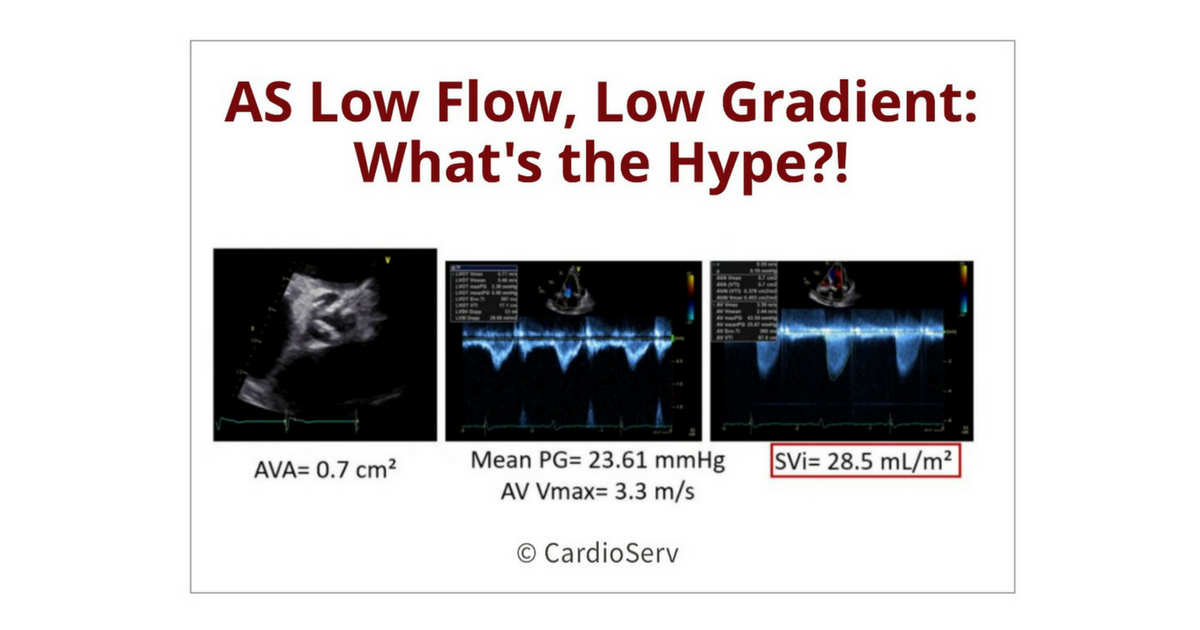
Aortic Stenosis Low Flow Low Gradient What S The Hype
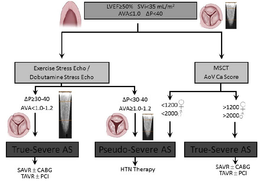
Complex Scenarios Paradoxical Low Gradient As In Normal Patients
Thevalveclub Com Br

Low Flow Low Gradient Aortic Stenosis When Is It Severe American College Of Cardiology

Prognosis Of Severe Low Flow Low Gradient Aortic Stenosis By Stroke Volume Index And Transvalvular Flow Rate Jacc Cardiovascular Imaging
Low Flow Low Gradient Aortic Stenosis When Is It Severe American College Of Cardiology
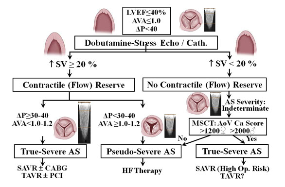
Complex Scenarios Low Gradient In Low Ef As Patients

Low Flow Low Gradient Aortic Stenosis When Is It Severe American College Of Cardiology